History of Radiography
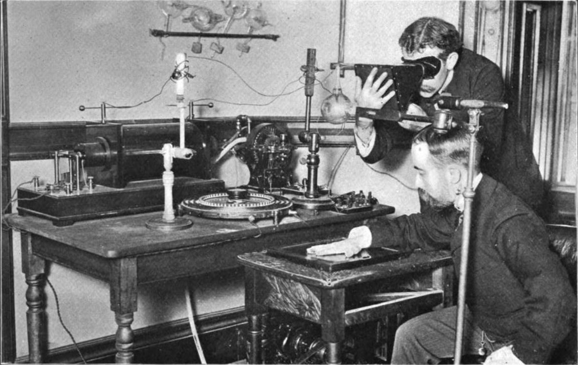
The individual saw for setting up radiography as known in bleeding edge is Wilhelm Conrad Roentgen, a German instructor. His examination focus work induced presentation of a ‘X-shaft’ that could experience huge paper, offering motivation to feel imperfect about shadows solid things. In a matter of seconds a short time later, Wilhelm would then apply his beginning late found exposure to human body organs, encouraging the first X-bar known on his wife’s hand.
The presentation had a tremendous impact on inspectors all around then. They quickly started changing the trial, beginning new fields of study identifying with this charming sort of segment. Individuals when all is said in done took huge excitement for general in the X-bar, with distinctive media outlets of the time spreading its news the entire course over their known world. As sad flimsiness remains, the disclosure would at last change both therapeutic structures and mechanical society for youngsters.
The 1,000,000-Volt X-Ray Generator
Radiography advanced gigantically after presentation of a 200,000-volt X-bar tube in 1922. This instrument empowered radiographs of thick steel areas to be put aside a couple of minutes periods. General Electric Company, in 1931, made X-bar generators of 1,000,000 volts, which changed into an influencing current photography contraption. American Society of Mechanical Engineers, ASME, that same year permitted utilization of X-bar machines for getting a handle on mix welded weight vessels. This further opened the course for unmistakable relationship to use and utilization radiography.
Deviousness by Radiation
Negative effects of X-sections discovered the chance to be evident as specialists broadened more imperative interest and upgraded each one of them over the globe. The people who worked with radiation reliably soon made certifiable wellbeing concerns from introduction to paying little heed to what you look like at it radiation. Inspectors in like way started considering how radiation had negative results for the body at cell level. Such research has requested that wellbeing physicists today show levels of radiation honorable for recuperating, present day and sensible applications.
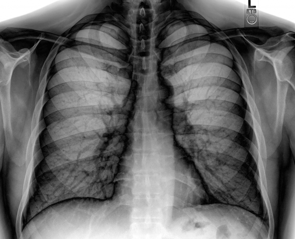
Radiography Applied at Present Day
Progress has made it possible to use radiography more than a colossal cross-extent of business attempts. Radiography is really used other than related enthusiastically outside the field of pharmaceutical. Radiography is indispensable for assessing things like airbags and canned food things. Additionally, it is used as a piece of systems for security at plane terminals near to specific workplaces. The radiography being utilized today is isolating for helping masters look at wellbeing issues which are not discernable physically within their patients. Radiography attracts forces to handle finding of tumors, mellowed bones and up expansion other wellbeing challenges with improved precision. New advances and PCs are instilled perpetually with radiographic routines for upgrading the quality and clarity of pictures passed on, relentlessly creating precision of therapeutic examination.
Studies in Radiography
Specialists and understudies are beginning now selecting for courses and classes in radiography. They discover the chance to take in the cutoff points required for adding to this field, among them how to work radiography conform in safe way. Certain radiography courses require an externship that allows understudies to couple hypothetical learning with hand-on relationship in their field of practice. Radiography has a phenomenally promising and goliath future in the healing associations industry. Scientists and supervisors are in a matter of seconds encouraging to push radiography and update considered patients through its application. The field of Radiography is turning out to be enthusiastic, with masters reckoning it is sure to continue influencing how the social certification structure investigate torments for all that much quite a while to come.