


Headway of Radiography Saving lives with X-Rays
Progression of Radiography Saving lives with X-Rays
X-columns are a kind of electromagnetic radiation like radio waves and light waves, next to their more lifted measures of hugeness, whereby they can enter the body. They first were discovered over a century before and discovered brief application in medicinal diagnostics. X-shafts remain a basic device today in performing conclusion adjacent treatment of different contaminations and wounds, the risks included in any case. All around, the approach which utilize X-columns for radiography join radiography, fluoroscopy, figured tomography (CT) yield, isotope or atomic prescription compass, PET and PET CT check.
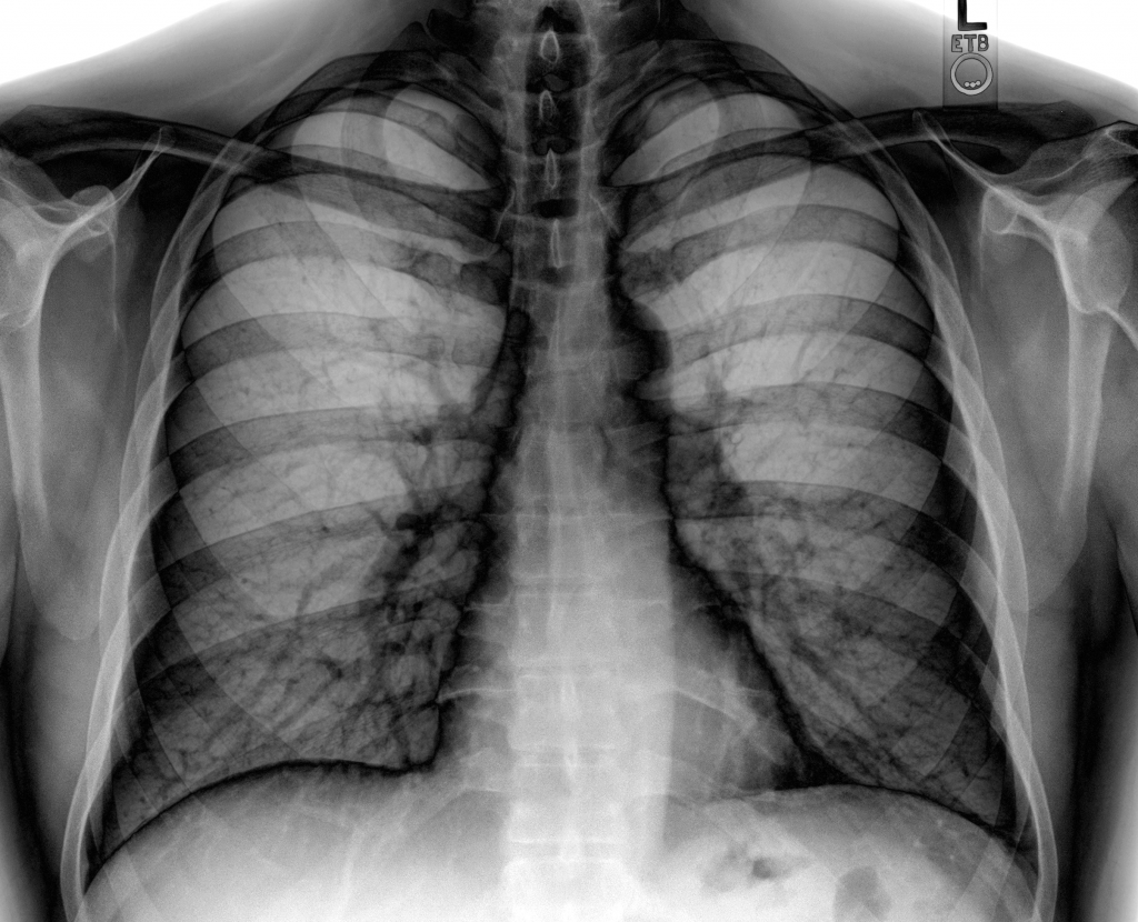
Radiography
This suggests the standard utilization of X-shafts. A X-bar machine makes a light outpouring columns, overseeing them to a touch of the body under examination and onto some marvelous film, enclosing a photo. A bigger bit of people have beginning now gotten a handle on dental X-columns, which are generally protected, low-estimation tests. X-bars are a significant part of the time performed on the midsection and broken bones, while mammography is an important test particularly for ladies that sees midsection infirmity in its starting stages. The tests use short measures of X-columns and in this manner just act little hazard to patients.
Fluoroscopy
The game plan of fluoroscopy uses X-shafts for conveying a moving picture on TV screen. “Still”, individual pictures may be picked and spared or the part spared altogether. This informative framework is valuable for exploring the guts or securing pictures of blood spilling inside of veins. A valid example, barium show in a barium supper is utilized for giving moving photos of the digestive tract and stomach. Iodine-based shading may be saturated into course to catch pictures of veins or the leg or heart, in a framework known as angiogram. This structure should be asked for of facilitating treatment frameworks like nephrostomy, which is spillage of blocked kidney, or angioplasty which infers amplifying of limited supply courses. Fluoroscopic examination ordinarily consolidates higher estimations of radiation rather than essential radiography.
Enrolled Tomography (CT) Scan
An enrolled tomography or CT extension is a to some degree complex methodology for applying X-columns, whereby the patient first lies on obliged table that encounters some meandering gap at purpose of union of the scanner. An impressive number of little X-column shafts enter a body-cut and arrive on banks of markers. Both the wellsprings of X-columns and pioneers make round turns inside of the machine. A PC shapes a photo of the cut, which is then shown by a TV screen. The patient passes the fissure a tiny bit at a time to catch pictures from assorted purposes of body and even make 3D photos every so often.
Isotope or Nuclear Medicine Scan
Isotope or Nuclear pharmaceutical yield is another radiographic framework in which a little fragment of radioactive material (isotope) gets embedded into the vein of a patient under examination. Then again, the material may once in a while be taken in or gulped. The radioactive material collects in a given tissue or organ, similar to the skeleton for bone yields.
PET range and PET CT Scan
This extraordinary kind of atomic arrangement compass perceives radiation from positron discharges. Positrons imply little particles which a radioactive substance transmits which then get controlled onto a patient. They can give data relating to how the heart or cerebrum limits or in perceiving contamination inside of the body.